
Philips expands network of clinical partners to set new standard of care for the early diagnosis and treatment of lung cancer Leave a comment
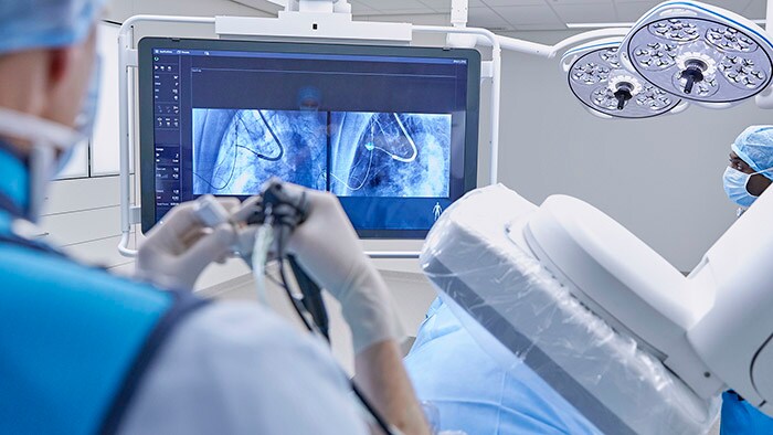
Philips Lung Suite provides advanced real-time 3D imaging with augmented fluoroscopy on the company’s Image Guided Therapy Systems – such as Azurion, combined with dedicated software. With Philips’ Cone Beam CT imaging, the X-ray detector rotates around the patient to generate a CT-like image in around five seconds, providing clinicians with a high-resolution 3D view of the target lesion and other anatomical structures. This allows the clinician performing the biopsy procedure to be continually guided by high-quality real-time imaging to advance a catheter towards the lesion through a bronchoscope. Once done, its position can be confirmed in real-time using the same imaging modality, and a biopsy sample can be taken.
“In the fast-growing world of intraoperative imaging, cone-beam CT remains the gold standard for augmented fluoroscopy and lesion confirmation. Using Lung Suite, no nodule can hide, regardless of anatomical position or radiologic characteristic, making it a valuable tool for both diagnosis and future ablation procedures,” said Dr. Amir Abramovich, MD., Director of Interventional Pulmonology at the Carmel Medical Center in Haifa, Israel.
“Cone-beam CT is the critical step towards targeting sub-20 mm nodules and an essential tool for the transition towards bronchoscopic microwave ablation of peripheral lung lesions,” said Professor Shah Pallav, MD., consultant respiratory physician at Royal Brompton Hospital in London, UK.
“The advanced cone beam CT imaging combined with augmented fluoroscopy of Philips Lung Suite gives us the confidence to safely reach and biopsy difficult-to-access peripheral lung nodules,” said Maarten Criel, MD., Pulmonologist at ZOL Genk Medical Center, Belgium.
Dr. Kelvin Lau, consultant and lead thoracic surgeon at St Bartholomew’s Hospital, London, successfully diagnosed and treated lung cancer patients using cone beam CT for biopsy, bronchoscopic microwave ablation and for precision image-guided surgery, all in one procedure, during initial clinical trial. Advanced imaging with Philips Lung suite is used during procedures for real-time 3D image guidance and confirmation. Additional clinical trials at various hospitals are expected to start soon.
An October 2021 paper in the Journal of Bronchology & Interventional Pulmonology authored by researchers at Radboud University Medical Center, the Netherlands, demonstrated the diagnostic potential of Lung Suite. The researchers were able to raise the diagnostic accuracy of navigation bronchoscopy from 72% to 90%. In addition, the average total effective radiation dose per procedure was reduced by more than half [3].
[1] https://www.who.int/news-room/fact-sheets/detail/cancer
[2] https://www.cancer.org/cancer/lung-cancer/detection-diagnosis-staging/survival-rates.html
[3] Journal of Bronchology & Interventional Pulmonology: October 2021 – Volume 28 – Issue 4 – p 262-271 doi: 10.1097/LBR.0000000000000783